|

|
|
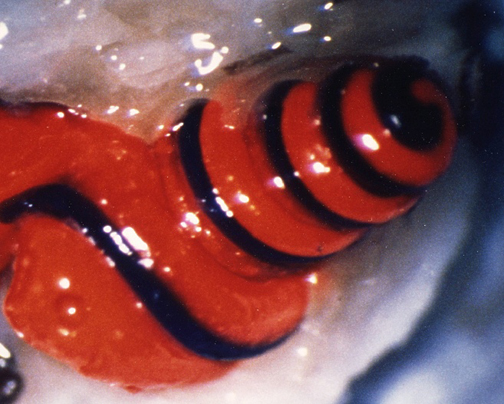
This image shows the two fluid spaces of the cochlea, the perilymph shown
orange and the endolymph shown blue. For years it was believed that endolymph
and perilymph flowed along the fluid spaces. We developed a method to measure
the flow rate in the living ear. The method used an ionic marker which was
iontophoresed into the fluids at low concentration. The concentration of the
marker was measured simultaneously by two ion-selective microelectrodes at
locations "upstream" and "downstream" of the injection site. Analysis
involved simulations of the combined effects of flow and diffusion along the
spaces.Ê We were able to show that the rate of flow of these fluids is
extremely slow, with endolymph flow shown to be less than 1 nanoliter per
minute. The method is extremely sensitive and detects very slow flow rates in
small compartments. For further details please CLICK
HERE.
Contributor: Prof.
Alec N. Salt, Department of
Otolaryngology, Washington University School of Medicine, St.
Louis, MO.
|
|